
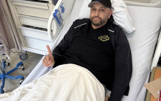
Becca Valle, experiencing severe headaches despite a seemingly content life, was initially puzzled. Living happily with her boyfriend and nearing family, Valle achieved a major personal milestone by running a marathon, reducing her stress levels. Yet, in September 2021, her headaches intensified to the point of debilitation. She suspected migraines, while her doctor considered sinus issues.
However, the situation escalated when Valle experienced excruciating pain, preventing her from sitting up or stopping vomiting. Promptly rushed to the hospital by her boyfriend, tests revealed bleeding in her brain, prompting an exploratory craniotomy. This procedure, involving partial skull removal, aims to examine the brain.
I wasn’t fully aware of the situation, which might have been a blessing. If I had realized the gravity of having my skull opened, I don’t think I would have coped well,
Valle, then aged 37, reflected.
During surgery, doctors discovered a brain tumor, which was removed in a subsequent craniotomy. Tests confirmed the tumor as glioblastoma. This type of cancer proves challenging, as complete surgical removal is rarely feasible. Even when tumors are entirely excised, like in Valle’s case, patients undergo chemotherapy and radiation to eliminate any residual cancer cells. The prognosis remains grim, with only about 10% of patients surviving beyond five years, according to the University of Texas’ MD Anderson Cancer Center.
Overcoming the Blood-Brain Barrier in Treatment
Treating glioblastoma is further complicated by the blood-brain barrier, a safeguarding cell layer limiting medication entry into the brain. While beneficial in reducing side effects, this barrier hinders effective brain-targeted treatments.
For Valle, joining a clinical trial exploring a method to temporarily open this barrier was a straightforward choice. It was a no-brainer. This could save my life,
Valle remarked.
The study, led by Dr. Graeme Woodworth, the chief of neurosurgery at the University of Maryland Medical Center, investigated whether focused ultrasound could momentarily breach the blood-brain barrier, enhancing chemotherapy’s effectiveness. This approach relied on sound waves interacting with microbubbles, typically used in imaging tests, which expanded and induced blood vessel permeability, allowing chemotherapy access to the brain.
Valle joined 34 participants undergoing three to six focused ultrasound treatments over six months, alongside routine MRIs to monitor cancer progress and any potential side effects. This innovative treatment necessitated wearing specialized headgear.
Encouraging Outcomes from the Trial
Trial data revealed a promising survival advantage for participants. Dr. Woodworth explained that while drug localization within the body wasn’t tracked during the trial, subsequent follow-ups highlighted patient longevity; 40% of trial participants survived longer than anticipated compared to a control group.
The control group, significantly larger than the study population, emphasized a survival advantage
for those combining focused ultrasound with oral chemotherapy compared to chemotherapy alone. Dr. Woodworth noted that these findings suggest that the monthly treatment is safe and potentially enhances survival and tumor control.
Dr. Patrick Wen of the Dana Farber Cancer Institute, although uninvolved in the trial, acknowledged the study’s significance, urged for broader research with varied chemotherapy regimens, and recommended randomized control groups for more robust data.
Dr. Woodworth’s team continues to seek validation and expansion of their research, though the University of Maryland’s current clinical trials focus on using the technique for treating brain metastases in specific lung cancer patients, pending FDA approval.
Exploring New Frontiers in Cancer Monitoring
Beyond treatment, the trial also fostered new research avenues. Pre- and post-treatment blood tests indicated alterations, suggesting potential for liquid biopsies to non-invasively monitor brain cancer progression. A secondary clinical trial conducted by Dr. Woodworth and team, awaiting FDA review, aspires to develop a practical device offering this capability.
Introducing this device into clinical settings could revolutionize treatment collaboration among doctors, pharmaceutical companies, and biotech firms,
shared Dr. Woodworth. An FDA-approved device capable of safely opening the blood-brain barrier could spark new effective treatment combinations for glioblastoma patients.
Becca Valle: A Story of Hope and Resilience
Despite having no trial side effects, Valle embodies a remarkable survivor story—four years post-diagnosis, her scans show no signs of disease despite the bleak statistics of glioblastoma.
I am truly an outlier,
expressed Valle, now 41. Valle, returning to her vibrant self, plans ski trips to Europe and embraces the present over statistics, needing only semi-annual MRIs. I don’t think about cancer. Worrying is unproductive. I’ve always embraced living life to the fullest—why change now?
she concluded.
 Cancer Death Rates Drop: Challenges in Addressing Disparities
  A Study on COVID-19 Vaccine Effectiveness Published
  Efforts to Combat Hospice Fraud and Protect Patient Care
  Study on Vaccine’s Impact on Emergency Visits and Hospitalizations
  Protein Tubulin Offers Hope in Combatting Alzheimer’s and Parkinson’s Disease
  Medicare Initiative Set to Lower Cost of Popular Weight-Loss Medications