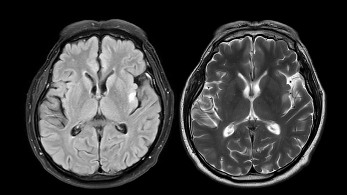
A brain scan tool measuring shrinkage in areas linked to thinking and memory may help predict the transition from mild cognitive impairment (MCI) to dementia in early-onset Alzheimer’s disease, a study suggests. This form of Alzheimer’s develops before age 65, often starting with MCI where memory and thinking problems are noticeable but not yet dementia.
People with early-onset Alzheimer’s progress at different rates from MCI to dementia. Researchers aimed to see if brain shrinkage patterns could indicate progression speed. They developed the early-onset Alzheimer’s disease signature, a tool to detect shrinkage in specific brain areas involved in thinking and memory, particularly parts of the parietal and temporal cortex.
Using MRI scans, researchers assessed the thickness of these areas, creating a score to represent brain shrinkage. The study followed 130 individuals with MCI who lived independently, and 97 healthy participants aged 40 to 64, over an average of two years. Around 65% with early-onset Alzheimer’s progressed from MCI to dementia, with greater gray matter shrinkage linked to faster progression. Each standard deviation increase in shrinkage upped the transition risk by 1.24 times.
This biomarker tool proved more effective in predicting progression than initial symptom analysis. Alexandra Touroutoglou from Harvard Medical School noted that many patients ask: “When will I lose my independence?” Current methods are limited in predicting progression from MCI to dementia in early-onset Alzheimer’s, but the study’s findings could help doctors estimate dementia onset timing.
Dr. Ronald Schwartz of Continuum Health Services explained that a reliable tool for forecasting progression can aid in setting expectations and support care planning. While acknowledging potential benefits in anticipatory guidance, Schwartz stressed that the tool should enhance discussions, not act as a definitive predictor.
The biomarker works as a map, pinpointing brain regions where shrinkage is more common in early-onset Alzheimer’s patients. Applied to MRI scans, it measures shrinkage to estimate progression speed. The study focused on a single participant group, primarily non-Hispanic White, so its applicability to diverse populations remains uncertain.
Further research must verify its accuracy across varied ages, stages, and clinical presentations. Precision in predictions could lead to improved care and patient-family decision-making. The findings imply that MRI-based gray matter shrinkage measurement may aid in planning, though broader testing of the tool is necessary.
Reference: Paranhos, T., et al. (2026). EOAD-Signature Atrophy Predicts Dementia in Early-Onset MCI due to Alzheimer Disease. Neurology. DOI: 10.1212/WNL.0000000000218519.
 Concerns Over Food Safety and Its Impact on Families
  A Mother’s Tragic Journey Under Abortion Ban
  Medical Diagnosis Errors: Understanding the Crisis
  Lymphatic Drainage and Ice Sculpting: Emerging Wellness Trends
  The Rising Trend of GLP-1 Medications Among College Students
  The Challenges and Opportunities of Testosterone Therapy for Menopausal Women